Ultrasound is a prenatal test that is offered to most pregnant women. This pregnancy scan uses sound waves to generate the picture of the baby inside the womb. Ultrasound is statutory for expecting mothers as it is the only diagnostic modality to measure the development of the fetus. Women with low-risk, complication-free pregnancies will typically have at least two ultrasounds, while older moms and those with complications will usually have more.
There are many reasons ultrasounds in general are necessary during pregnancy, depending on the trimester, including:
The Wide Network of Best Healthcare
Our team of highly trained professionals uses the latest healing technologies to restore you to pain-free health quickly and easily.
The Wide Network of Best Healthcare
Our team of highly trained professionals uses the latest healing technologies to restore you to pain-free health quickly and easily.
Types of Ultrasound Examinations
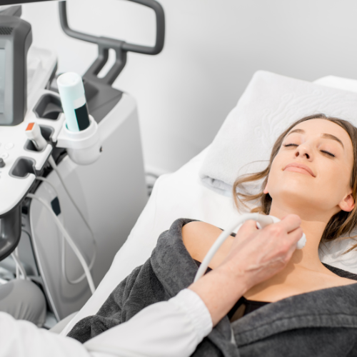
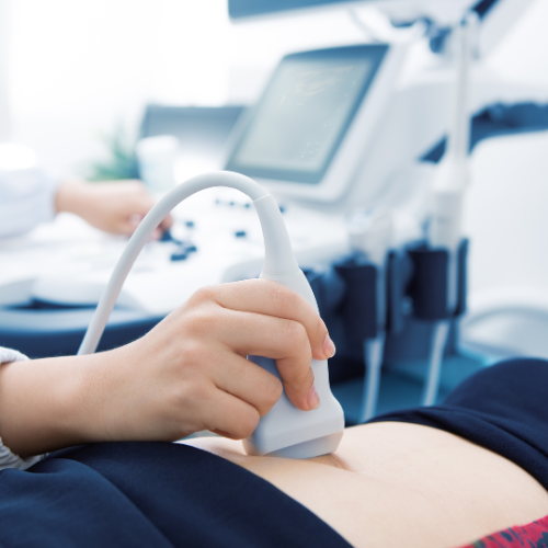
Transabdominal Ultrasound
The most common type of prenatal ultrasound, it involves applying gel on the mother's abdomen and using a handheld device (transducer) to capture images by moving it over the belly. This method is generally used for routine prenatal screening and monitoring.
Doppler Ultrasound
Doppler ultrasound uses sound waves to assess the blood flow the fetus, placenta, or umbilical cord. It helps evaluate fetal well-being and detect any abnormalities in blood circulation.
3D Ultrasound
The Ultrasound is used to take numerous pictures at a time. It creates a 3D image.
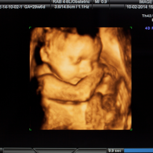
4D Ultrasound
The Ultrasound is like the 3D ultrasound but additionally, it shows the movement of the baby in form of a video.
Anomaly
A 20-week ultrasound, sometimes called an anatomy scan or anomaly scan, is a prenatal ultrasound performed between 18 and 22 weeks of pregnancy.